Figure 2.
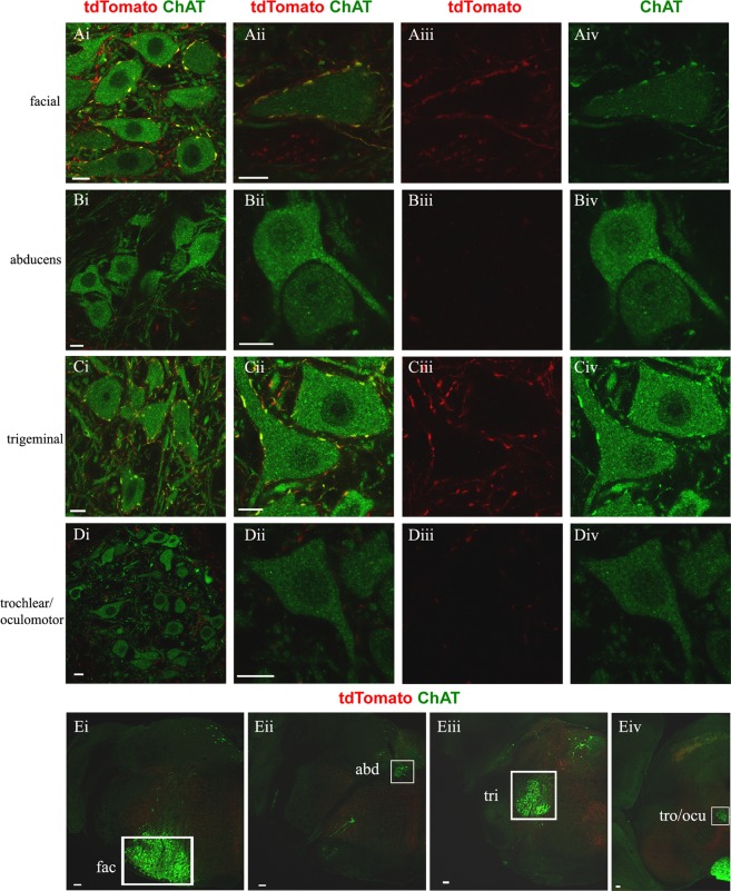
C boutons on brainstem motor neurons are Pitx2 derived. In green anti-ChAT antibody labels motor neuron somata and cholinergic C boutons on them, in red anti-dsRed antibody labels tdTomato+ Pitx2 derived synapses. (A–D) The first column contains images of the cranial motor nuclei and the next three columns contain close up images of motor neurons (merge, red only, green only). (Ai–iv) Pitx2 derived (red) C boutons (green) on the facial nucleus (green) appear yellow. (Bi–iv) Nucleus abducens does not receive C bouton inputs. (Ci–iv) Pitx2 derived (red) C boutons (green) on the trigeminal nucleus (green) appear yellow. (Di–iv) Trochlear and oculomotor nuclei do not receive C bouton inputs. Scale bar (A–D) 10 μm. (Ei) Half section of medulla showcasing the position of facial nucleus, (Eii) nucleus abducens, (Eiii) trigeminal nucleus, (Eiv) trochlear/oculomotor nucleus, in white boxes. Scale bar (E) 100 μm.

