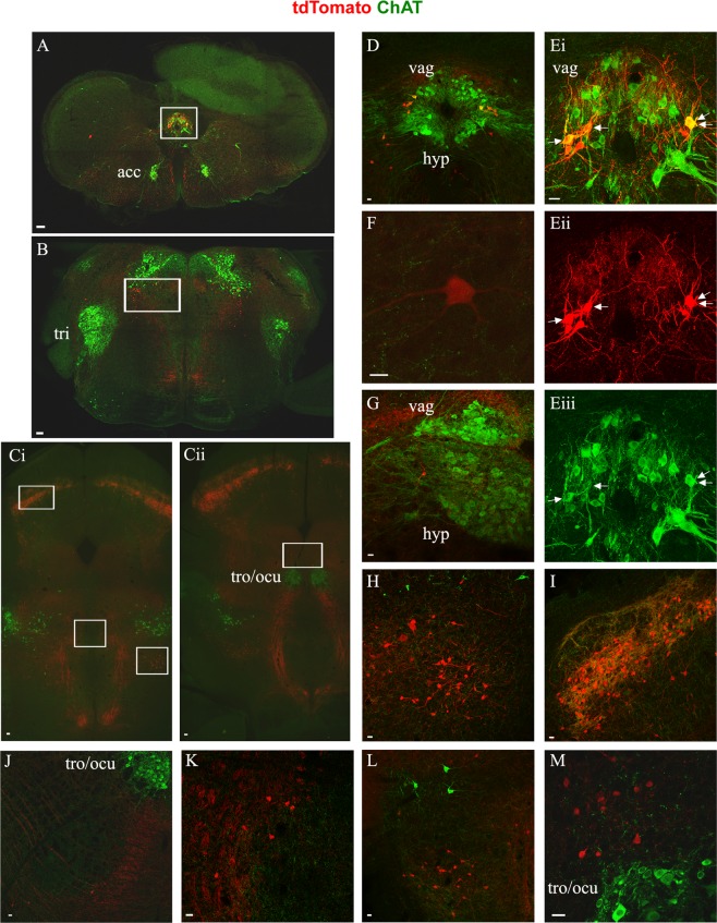
Figure 4.
Pitx2 neurons of the brainstem. In green anti-ChAT antibody labels motor neuron somata, cholinergic interneurons and cholinergic synapses from interneurons and motor neurons, in red anti-dsRed antibody labels tdTomato+ Pitx2 neurons and Pitx2 derived synapses. Sections (A–C) presented in caudal to rostral succession (A) Caudal medulla section showcasing the localization of Pitx2 cholinergic and non-cholinergic neurons, in white box. (B) Pons section at the level of trigeminal nucleus. In red Pitx2 non-cholinergic neurons ventromedial and ventral of the laterodorsal tegmental nucleus indicated in white box. (Ci,ii) Midbrain sections, i. in white boxes the populations of the superior colliculus, of the anterior tegmental nucleus and of the penduculopontine tegmental nucleus, ii. in white box the population in the supraoculomotor area. Scale bar (A–C) 100 μm. (D) Close up image of the caudal medulla Pitx2 neurons among the dorsal nucleus of vagus and the hypoglossal. (Ei–iii) Close up image of caudal medulla cholinergic Pitx2 neurons, indicated with white arrows (i. merge, ii. red only, iii. green only). (F) Occasional non-cholinergic Pitx2 neurons appear in a lateral position away from the central canal. (G) The most rostral Pitx2 neurons in the medulla appear approximately in the middle of the dorsal nucleus of vagus and hypoglossal nucleus in the rostrocaudal axis. (H) Close up image of pons Pitx2 neurons. (I) Close up image of the Pitx2 neurons in the superior colliculus among cholinergic axons and synapses. (J) Close up of Pitx2 neurons in the red nucleus. (K) Close up of the Pitx2 neurons around the anterior tegmental nucleus (ATg). (L) Close up of Pitx2 neurons in the oral part of the pontine reticular nucleus (PnO), lateral to the tectospinal tract (ts) ventral to the penduculopontine tegmental nucleus (PPTg). (M) Close up image of Pitx2 neurons in the supraoculomotor area Scale bar (D–K) 20 μm.

