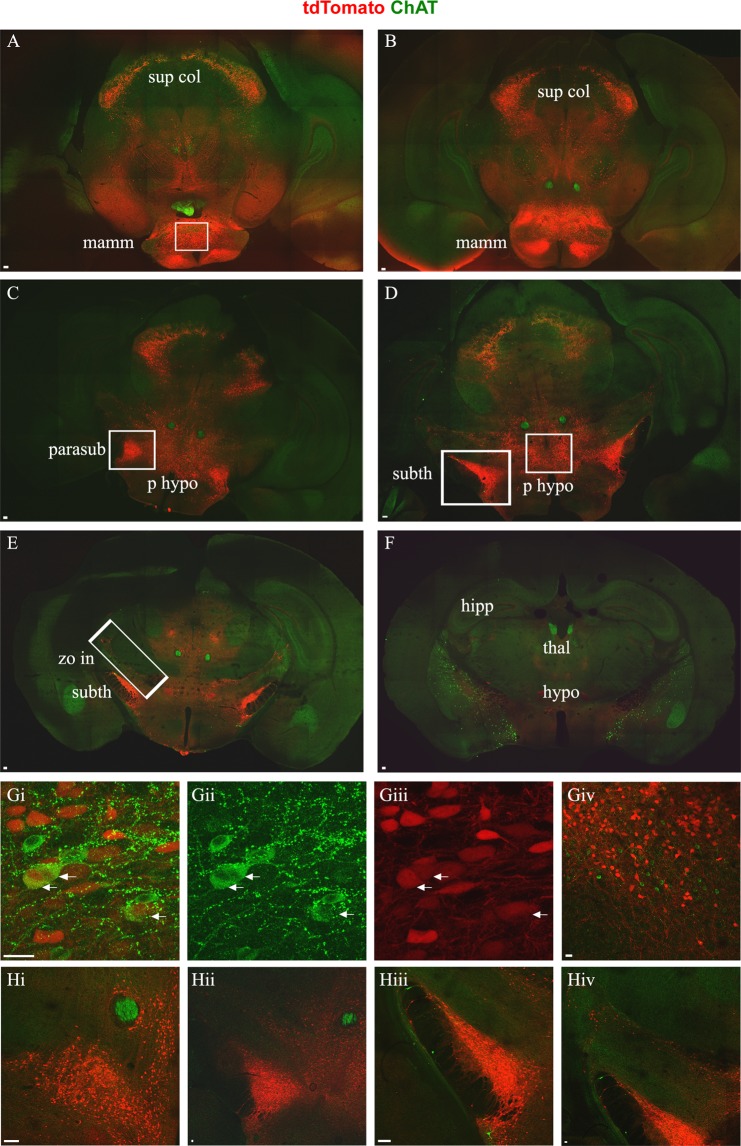
Figure 5.
Pitx2 neurons of the anterior brain. In green anti-ChAT antibody labels motor neuron somata, cholinergic interneurons and cholinergic synapses from interneurons and motor neurons, in red anti-dsRed antibody labels tdTomato+ Pitx2 neurons and Pitx2 derived synapses. Sections (A–F) presented in caudal to rostral succession. (A) Pitx2 neurons in the mammillary area of the hypothalamus are present in the ventral part of the section (white box). The vast majority are non-cholinergic and very few at the boundary of supramammillary and medial mammillary nucleus are cholinergic. At the dorsal part of the section the midbrain Pitx2 neurons are still evident. (B) More rostral section with Pitx2 neurons in the mammillary area. (C) Pitx2 neurons evident in the posterior hypothalamus in the medial part of the section and the parasubthalamic nucleus at the lateral sides (white box). (D) At the lateral sides of the section, Pitx2 neurons appear in the subthalamic nucleus and in the medial area in the posterior hypothalamus (white boxes). (E) Subthalamic Pitx2 neurons and a line of Pitx2 neurons in the dorsal part of the zona incerta, dorsally to subthalamic nucleus (white box). (F) More rostral section that lacks Pitx2 somata, but few Pitx2 axons are still present. Scale bar (A–F) 100 μm. (Gi–iii) Close up image of cholinergic mammillary Pitx2 neurons indicated with arrows (i: merge, ii: green only, iii: red only). (Giv) Close up image of the mammillary area with non-cholinergic Pitx2. (Hi) Close up image of the non-cholinergic Pitx2 neurons in the posterior hypothalamus. (Hii) Close up image of the non-cholinergic Pitx2 neurons in the parasubthalamic nucleus. (Hiii) Close up image of the non-cholinergic Pitx2 neurons in the subthalamic nucleus. (Hiv) Close up image of the non-cholinergic Pitx2 neurons in the dorsal part of zona incerta, dorsally to the subthalamic nucleus. Scale bar (G,H) 20 μm.

