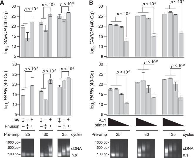
Figure 3.
A blend of Taq–Phusion polymerases improves selective poly(A) amplification of cDNA and reduces AL1 primer requirements. Cells were obtained by LCM from a human breast biopsy and split into 10-cell equivalent amplification replicates. (A) Poly(A) PCR was performed with 15 µg of AL1 primer with Taq alone (10 units), Phusion alone (4 units) or Taq/Phusion combination (3.75 units/1.5 units). (B) Poly(A) PCR was performed with either 25, 5, 2.5, or 0.5 µg of AL1 primer and the Taq–Phusion blend from (A). Above—Relative abundance for the indicated genes and preamplification conditions was measured by quantitative PCR (qPCR). Data are shown as the median inverse quantification cycle (40–Cq) ± range from n = 3 amplification replicates and were analysed by two-way (A) or one-way (B) ANOVA with replication. Below—Preamplifications were analysed by agarose gel electrophoresis to separate poly(A)-amplified cDNA from nonspecific, low molecular-weight concatemer (n.s.). Qualitatively similar results were obtained separately three times. Lanes were cropped by poly(A) PCR cycles for display but were electrophoresed on the same agarose gel and processed identically. The uncropped image is shown in Supplementary Fig. S13A.

