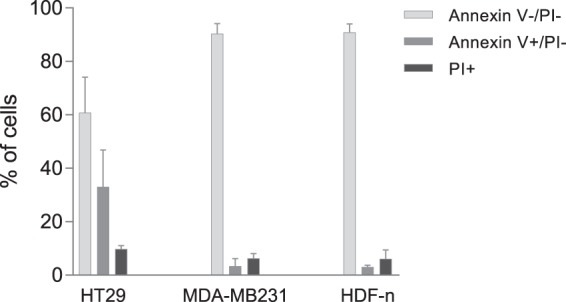
Figure 4.

Flow cytometric analysis of phosphatidylserine (PS) exposure. Percentage of PS-exposing cells in the outer leaflet of the plasma membrane in each cell line. Untreated cells were stained with propidium iodide (PI) and Annexin V before flow cytometric analysis. The Annexin V stain is much used to detect apoptosis; however here we investigated untreated cells under the assumption that the PS observed in the outer leaflet was not a result of apoptosis but related to the composition of the cell membrane. Thus, Annexin V+/PI− show the percentage of cells exposing PS in the outer membrane. PI+ show the percentage of dead cells. Mean + SD, n = 3–4, each performed as individual experiments.
