Figure 1.
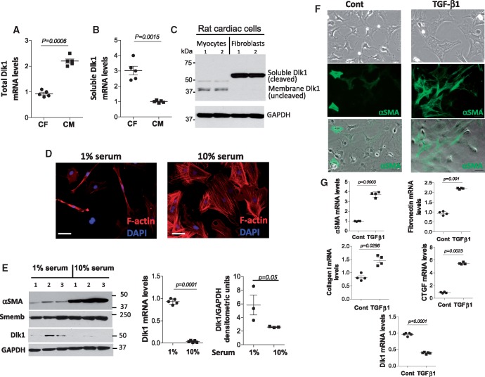
Cardiac cells express different delta-like homologue 1 (Dlk1) isoforms with delta-like homologue 1 expression decreasing in fibroblasts differentiating into myofibroblasts. Real time RT-PCR of total delta-like homologue 1 isoform (A) and delta-like homologue 1 soluble/cleaved (B) encoding mRNA variants from cardiac myocytes (CM) and fibroblasts (CF). (C) Representative western blot of delta-like homologue 1 reveals bands of different molecular weights corresponding to delta-like homologue 1 isoforms in cardiac myocytes and fibroblasts. GAPDH is a loading control. (D) Representative images of mouse cardiac fibroblasts maintained in 1% or 10% serum showing the effect on morphology and stress fibre expression (F-actin, red). (E) Western blot for α-smooth muscle actin, Smemb and delta-like homologue 1; GAPDH is a loading control. Graphs represent delta-like homologue 1 mRNA expression and protein densitometric analysis of delta-like homologue 1. (F) Representative images from non-treated (Cont) and treated (TGF-β1) fibroblasts cultured without serum. Top panel displays the change of fibroblast morphology after 24 h with TGF-β1. Middle (α-smooth muscle actin, green) and lower (merged) panels show an enhanced α-smooth muscle actin expression in TGF-β1-treated fibroblasts. (G) qRT-PCR of α-smooth muscle actin, fibronectin, collagen 1, connective tissue growth factor (CTGF), and delta-like homologue 1 in non-treated and TGF-β1-treated fibroblasts. Scale bars: 40 µm.

