Figure 2.
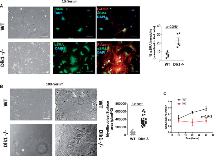
Lack of delta-like homologue 1 promotes fibroblast-to-myofibroblast conversion in cardiac fibroblasts. (A) Representative images exhibiting elongated wild-type fibroblasts vs. large, polygonal-shaped delta-like homologue 1-null fibroblasts cultured in the presence of 1% serum (left panel). Immunostaining of α-smooth muscle actin (middle panels, green) and F-actin (right panels, red) in wild-type and delta-like homologue 1-null fibroblasts with α-smooth muscle actin staining quantification (right panel; n = 5 different staining with 10–25 cells/field). Arrows point to co-localization of α-smooth muscle actin and stress fibres. (B) Images from wild-type and delta-like homologue 1-null fibroblasts cultured in the presence of 10% serum with fibroblasts lacking delta-like homologue 1 appearing large with a better organized actin cytoskeleton. Right panel shows cells at higher magnification with size quantification (n = 16 different fields with 5–10 cells/field. (C) Cell proliferation analysis assessed by incorporation of BrdU at 16, 20, and 24 h from wild-type and delta-like homologue 1-null fibroblasts. Scale bars: (A) 40 µm; (B) 40 µm (left images), 25 µm (right images).

