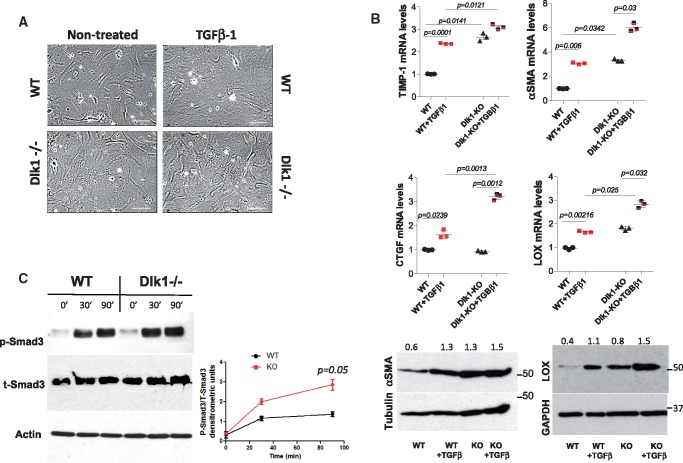
Figure 3.
Deletion of delta-like homologue 1 leads to activation of TGF-β1 signalling. (A) Representative images of wild-type and delta-like homologue 1-null fibroblasts ± TGF-β1 for 24 h (10 ng/mL). Scale bars: 40 µm. (B) qRT-PCR for TIMP-1, α-smooth muscle actin, connective tissue growth factor, and lysyl oxidase from wild-type and delta-like homologue 1-null fibroblasts ± TGF-β1 (upper panel) with representative western blots of α-smooth muscle actin and lysyl oxidase (lower panel). The numbers indicate α-smooth muscle actin and lysyl oxidase densitometric analyses normalized to tubulin and GAPDH loading, respectively. (C) Western blot analysis of phosphorylated Smad-3 in wild-type and delta-like homologue 1-null fibroblasts ±TGF-β1 for 30 and 90 min. Time 0 shows basal levels of phospho-Smad3. Graph represents the ratio of phospho-Smad3/total Smad3 at 0, 30, and 90 min.

