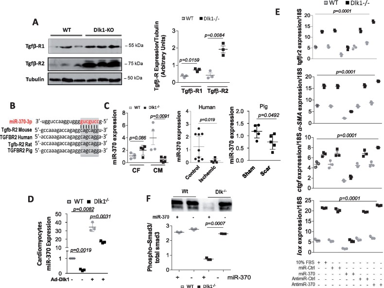
Figure 7.
Delta-like homologue 1 activates miR-370 and inhibits TGF-β1/Smad3 signaling. (A) Representative western blot analysis and densitometry quantification of TGFβ-R1 and 2 in wild-type and delta-like homologue 1-null hearts; Tubulin is a loading control. (B) Bioinformatics analysis identified TGFβ-R2 as putative target gene of miR-370. (C) qRT-PCR quantification of miR-370 in cardiac fibroblasts (CF) and cardiomyocytes (CM) isolated from wild-type and delta-like homologue 1-null mice, in healthy human and ischaemic patients, and in scar area in pigs vs. sham hearts. (D) qRT-PCR quantification of miR-370 following Ad.Dlk1 infection of cardiomyocytes (MOI100) from wild-type and delta-like homologue 1-null mice. (E) qRT-PCR quantification of TGFβ-R2, α-smooth muscle actin, connective tissue growth factor, and lysyl oxidase in fibroblasts isolated from Dlk1−/− or wild-type hearts treated with control miR (miR-Ctrl), miR-370 mimics (miR-370), control AntimiR (AntimiR-Ctrl), or miR-370 inhibitor/antimiR (AntimiR-370). 10% FBS is a positive control. Multiple comparisons were performed by analysis of variance (ANOVA) with post hoc Bonferroni’s test. (F) Western blot analysis and quantification of Smad3 phosphorylation (normalized to total Smad3) in wild-type and delta-like homologue 1-null fibroblasts ± miR-370 mimics.

