
FIGURE 1.
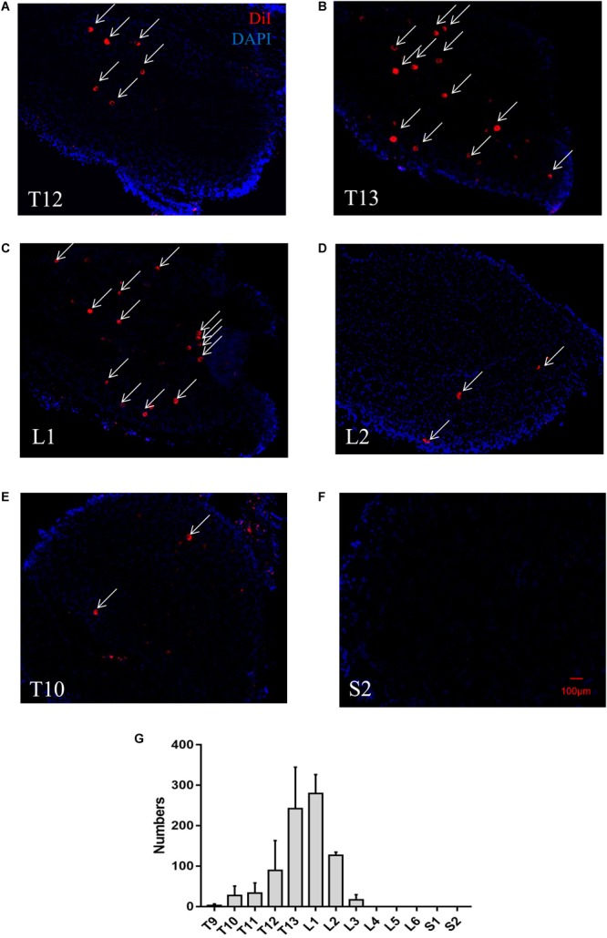
A representative retrograde trace of a DRG after DiI injection into perirenal adipose tissue (PrAT) in vivo. (A–D) Numerous DiI-labeled neurons (indicated by arrows) were found in the T12-L2 DRGs. (E) Few DiI-labeled neurons were found in the T10 DRG. (F) No DiI-positive neurons were found in the sacral DRG. (G) The numbers of PrAT-innervating neurons in DRGs throughout the spine. The largest numbers of neurons were found in the T13 and L1 DRGs.