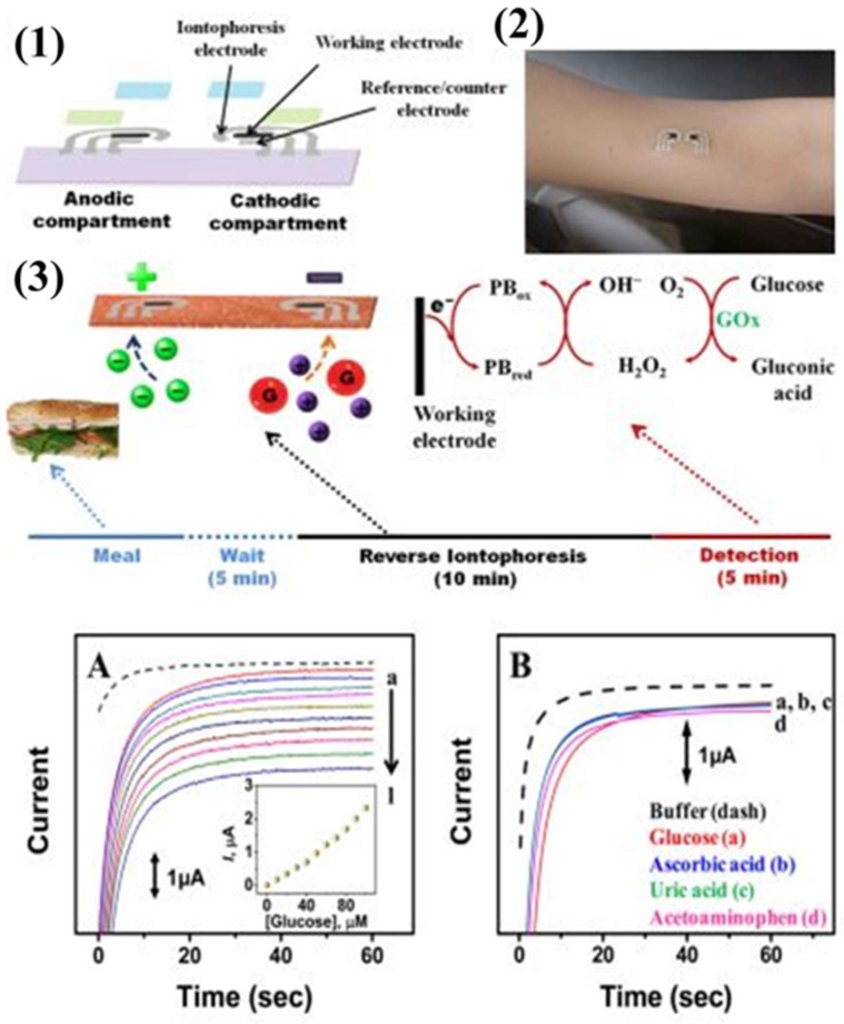
Figure 6.
Tattoo-based platform for noninvasive glucose sensing. (1) Schematic of the printable iontophoretic-sensing system displaying the tattoo-based paper (purple), Ag/AgCl electrodes (silver), Prussian Blue electrodes (black), transparent insulating layer (green), and hydrogel layer (blue). (2) Photograph of a glucose iontophoretic sensing tattoo device applied to a human subject. (3) Schematic of the time frame of a typical on-body study and the different processes involved in each phase. (A) Chronoamperometric response of the tattoo-based glucose sensor to increasing glucose concentrations from 0 μM (dashed) to 100 μM (plot “l”) in buffer in 10 μM increments. (B) Interference study in the presence of 50 μM glucose (plot “a”), followed by subsequent 10 μM additions of ascorbic acid (plot “b”), uric acid (plot “c”), and acetaminophen (plot “d”). Potential step to −0.1 V (vs. Ag/AgCl). Medium was phosphate-buffer with 133 mM NaCl (pH 7) [21].

