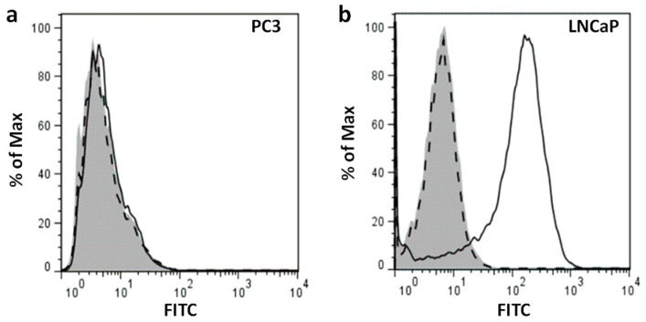
Figure 2.
Flow cytometry of PSMA expression in (a) PC3 and (b) LNCaP cells. Anti-human PSMA antibody conjugated to Alexa Fluor® 488 and corresponding isotype control were used to stain the cells. Unstained cells are shown in gray. Dashed lines indicate the isotype control and solid lines show PSMA-positive cells.

