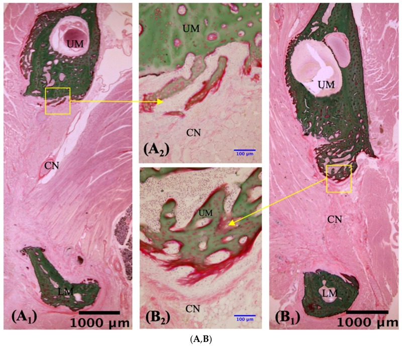
Figure 6.
Villanueva Goldner staining of undecalcified sections in the no-transplantation (n = 3), HBSS (n = 3), 1 × 10⁴ hMSCs (n = 3), and 1 × 10⁵ hMSCs (n = 3) groups. ((A,C,E,G); n = 3) Slices prepared 2 weeks after creation of the mandibular defect. ((B,D,F,H); n = 3) Slices prepared at 4 weeks. Newly formed bone was observed on and in the material shown in panels (E–H). RM, residual material; UM, upper mandible; LM, lower mandible; NB, newly formed bone; CN, connective tissue. The blue arrows in panels (F3,G3) indicate cuboidal osteoblast-like cells that lay in rows adjacent to newly formed bone. The green arrows in panels (E3,F3,G3,H3) indicate chondrocytes that synthesize the cartilaginous extracellular matrix. (A1,B1,C1,D1,E1,F1,G1,H1): ×1.25 magnification. (A2,B2,C2,D2,E2,F2,G2,H2): ×20 magnification. (E3,F3,G3,H3): ×40 magnification. Scale bars: 1000 μm (black), 100 μm (blue), 50 μm (red).


