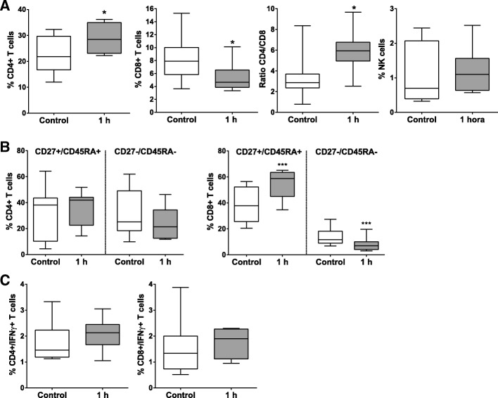
Fig. 1.
Lymphocyte subsets distribution in peripheral blood. Peripheral blood lymphocytes were isolated from blood samples collected before acute myocardial infarction model creation (control) and 1 h after it. The blood lymphocytes subsets (a). differentiation/activation T cell subsets (b) and IFNγ+ cells (c) were analyzed by flow cytometry. Paired comparisons were performed using a Student t-test for parametric data. Graphs show the mean ± SD (n = 11) of subpopulation subsets. * ≤ 0.05. ** ≤ 0.01. *** ≤ 0.001

