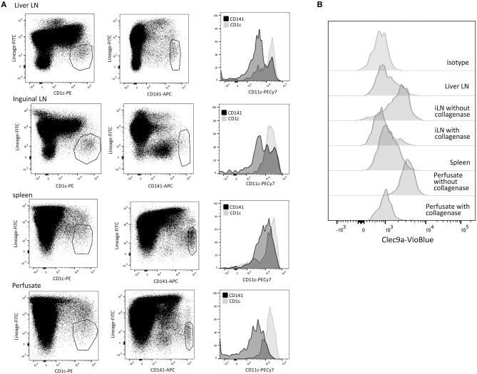
Figure 1.
Characterization of cDC subsets in hepatic and inguinal lymph nodes, spleen, and liver. (A) Vital (7-AAD−)CD45+Lineage−CD1c+ and CD45+Lineage−CD141+cells were gated in MNC isolated from hepatic and inguinal lymph nodes, spleen and liver perfusate, and analyzed for CD11c expression. (B) Vital (7-AAD−)CD45+Lineage−CD141bright cells were analyzed for Clec9A expression. Cells isolated from lymphoid tissues showed low Clec9A expression, while their counterparts in liver perfusate were Clec9A+. When liver perfusate cells were incubated with collagenase, Clec9A expression was lost. When leukocytes were isolated from inguinal LN without collagenase digestion, Clec9A was expressed on cDC2. iLN, inguinal LN.

