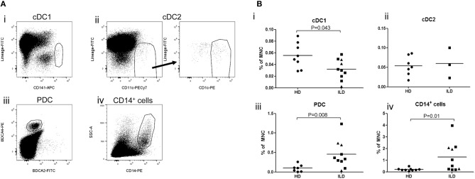
Figure 6.
(A) Relative numbers of APC subsets in liver lymph nodes of patients with inflammatory liver diseases. Flowcytometric analysis of cDC1 and cDC2 in single cell suspension of a LN attached to an explanted liver of a PSC patients. Within viable CD45+ cells, CD141hi cDC1 and CD11c+CD1c+ cDC2 were gated. (B)The percentages of cDC1, cDC2, PDC, and CD14+ cells were determined within CD45+ MNC in hepatic lymph nodes derived from healthy donors (HD; n = 8) and patients with inflammatory liver disease (ILD; n = 10 for cDC1 and n = 3 for cDC2). Patients with primary sclerosing cholangitis are depicted as squares and patients with auto-immune hepatitis as triangles. Lines indicate means.

