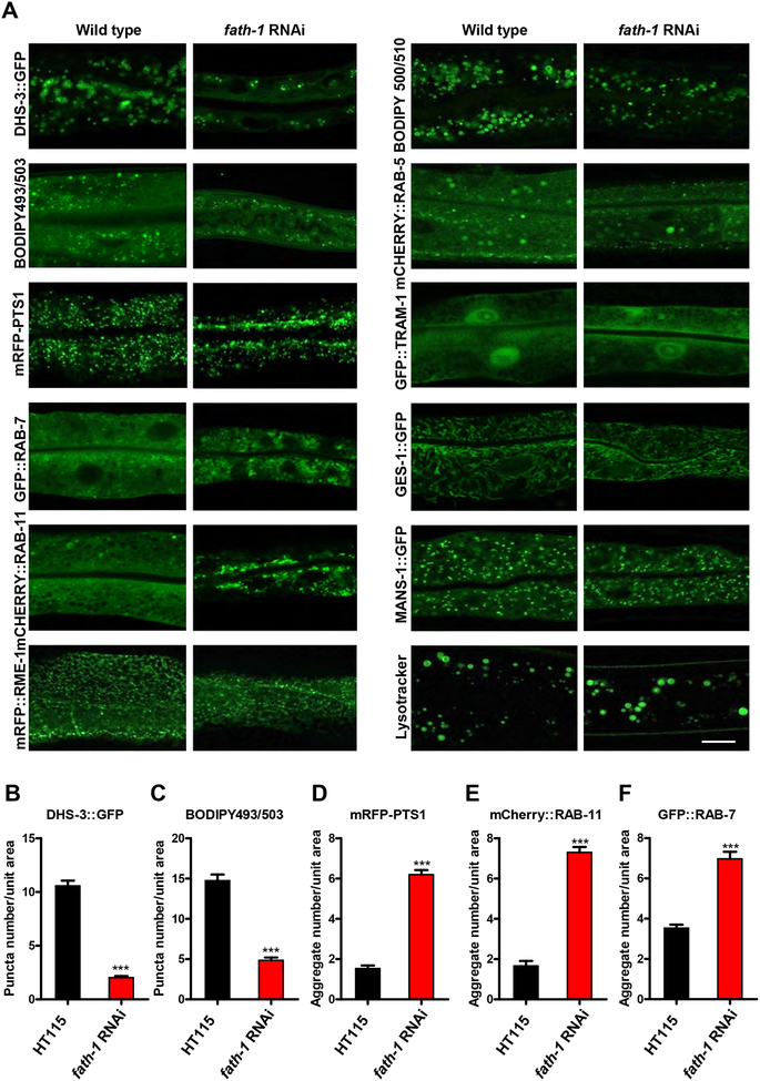
Figure 4. The subcellular defects caused by fath-1 inactivation.
(A) Representative fluorescent confocal images showing intestinal cellular organelles of young adult C. elegans treated with HT115 or fath-1 RNAi. The organelles include: lipid droplets (DHS-3::GFP, BODIPY 493/503); peroxisomes (mRFP::PTS1); late endosomes (GFP::RAB-7); apical recycling endosomes (mCherry::RAB-11); basolateral recycling endosomes (mRFP::RME-1); early endosomes (mCherry::RAB-5); endoplasmic reticulum (GFP::TRAM-1); mitochondria (GES-1::GFP) or Golgi (MANS-1::GFP). Lysosomes were marked by lysotracker green. BODIPY 500/510 visualizes the process of fatty acid uptake. The scale bar represents 10 μm. (B, C, D, E, F) Quantification of the numbers of endosome and peroxisome aggregates or lipid droplets in worms lacking fath-1 expression, corresponding to results shown in A. N ≥ 40 for each data set. Error bars, ± SEM; ***p<0.001.

