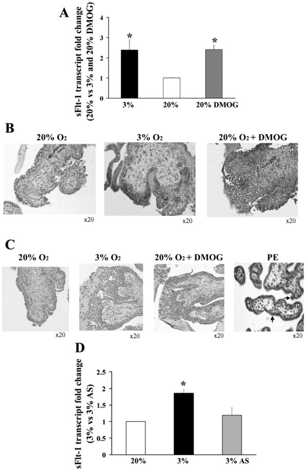
Fig. 5.
Effect of dimethyloxalyl-glycin (DMOG) and antisense oligonucleotides to hypoxia inducible factor (HIF-1α) on sFlt-1 expression in first-trimester placental explants. A: effect of DMOG treatment on sFlt-1 transcript in villous explants assessed by qRT-PCR, n = 3. *P < 0.05, 3% and 20% DMOG vs. 20% O2. B: effect of DMOG treatment on spatial localization of sFlt-1 protein in villous explants, n = 3. Dark gray staining represents positive immunoreactivity. C: TUNEL staining of villous explants and placental sample of early PE. Positive staining appears as black nuclear staining. D: effect of antisense oligonucleotides to HIF-1α (AS) on sFlt-1 mRNA expression in explants, n = 6. *P < 0.05, 3% O2 vs. 3% O2 +AS. Values are expressed as means ± SE of at least three separate experiments carried out in triplicate.

