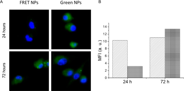
Figure 6.
(A) Fluorescence microscopy images of murine BMDCs incubated with FRET NPs and green NPs for 24 and 72 h collected at the green emission channel. Cell nuclei are stained with DAPI (blue). (B) Mean fluorescence intensity value BMDCs incubated with green NPs (light gray) and FRET NPs (dark gray) measured at the green emission channel using flow cytometry. A complete recovery of donor quenching is observed at 72 h.

