
Figure 5.
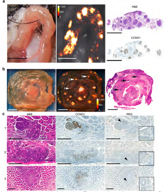
SERRS NP-mediated detection of intestinal adenomas. (a) In vivo Raman imaging was performed on an exteriorized bowel loop in an isoflurane-anesthetized ApcMin/+ mouse. Raman imaging identified multiple SERRS-positive lesions (middle panel; SERRS signal in counts per second) that all corresponded to adenomas as confirmed by histology (right panel; H&E staining and CCND1 immunohistochemistry). All scale bars: 5 mm. (b) To determine the sensitivity of SERRS-based Raman imaging for (pre-) malignant lesion detection, segments of the small intestine of ApcMin/+ mice (n = 4) injected with SERRS-NPs were scanned by Raman imaging ex vivo (middle panel; intensity in counts per second; scale bars, 5 mm). Even lesions less than 1 mm were detected by Raman imaging and would have been missed by macroscopic observation. The tissues were sectioned, and the 5 μm tissue sections were stained with H&E (right panel) and for CCND1 and PEG, respectively. (c) SERRS-positive lesions were correlated to histologic appearance and CCND1 status. As shown for the lesions indicated by arrowheads 1−3 in panel (b), SERRS signal correlates to the presence of dysplastic lesions. α-PEG was used to corroborate the presence of the SERRS-NPs (all scale bars, 500 μm; insets show 16× magnifications of the areas indicated by the arrow heads). See also Table 1 and Supplementary Figures S5−S7.