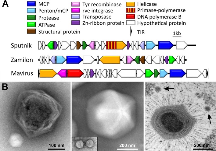
Fig 1. Genome organization and capsid shape of cultured virophages.
(A) Genome representation of the virophages Sputnik, Zamilon, and mavirus. Homologous genes are colored identically. (B) Electron microscopy images depicting capsids of giant viruses and their associated virophages. (Left) CroV (dark) and mavirus (light); negative stain EM courtesy of U. Mersdorf, MPI for Medical Research, Germany. (Middle) Megavirus vitis (with a visible stargate structure) and Zamilon vitis (inset); negative stain EM courtesy of C. Abergel, Aix-Marseille Université, France. (Right) Acanthamoeba polyphaga mimivirus with two Sputnik virus particles (arrows); thin-section EM courtesy of J.Y. Bou Khalil and B. La Scola, IHU Mediterranée Infection, France. Note that all three virophages have similar capsid sizes but are shown here at different magnifications. EM, electron microscopy; CroV, Cafeteria roenbergensis virus; TIR, terminal inverted repeat.

