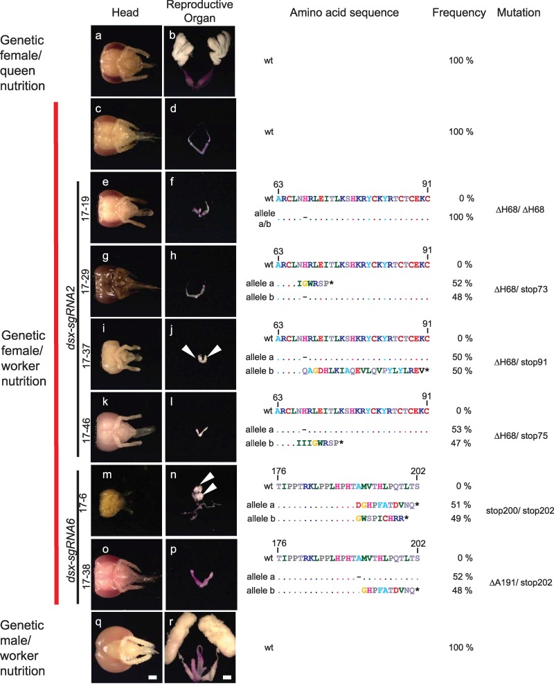
Fig 4. Size polyphenism of the reproductive organs in genetic female double mutants for the dsx gene.
Pictures of the head and internal reproductive organs of mutated and WT control bees are shown on the left, while the genotypes at the dsx locus with the deduced amino acid sequences are displayed on the right. Mutated and control genetic females and males were reared on worker nutrition. Queens were reared on the queen diet in a colony (we cannot mimic queen rearing in the laboratory). The WT amino acid sequence is shown above the detected alleles for comparison. (a, b) WT genetic female reared on queen nutrition (RJ) in the colony. (c, d) WT genetic females manually reared on worker nutrition. (e–l) Genetic females reared on worker nutrition that were double mutants for dsx via the dsx-sgRNA6 (note that a small part of the worker bee head 17–39 [picture i] is missing due to the dissection process). (m–p) Genetic females reared on worker nutrition that were double mutants for dsx via the dsx-sgRNA2. (q, r) Genetic males manually reared on worker nutrition. Organs were stained with aceto-orcein (reddish coloring) to facilitate the dissection process. Testis tissues are marked with arrows. Scale bar, 1 mm. Dashes in the sequence indicate deletions, and stars illustrate early translational stop codons. RJ, royal jelly; WT, wild type.

