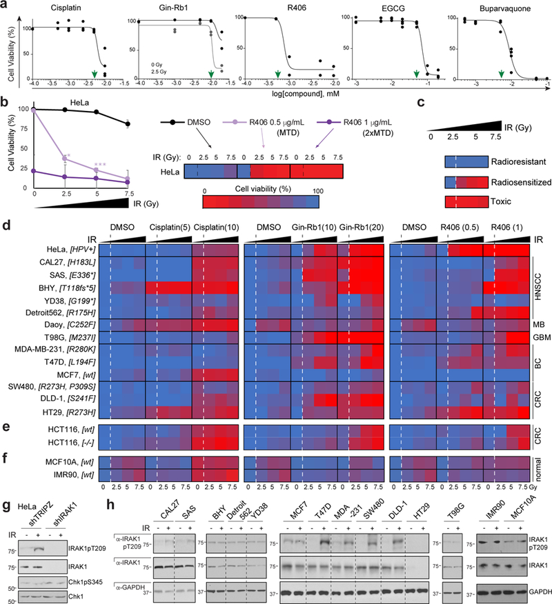
Fig. 5. IRAK1 inhibitors restore radiosensitivity across TP53 mutant tumour cell models.

(a) AlamarBlue-based HeLa cell viability assays establish MTDs (green arrowheads) for cisplatin, IRAK1 inhibitors Ginsenoside-Rb1 (Gin-Rb1) and R406, and PIN1 inhibitors EGCG and buparvaquone. Gin-Rb1 required 2.5 Gy to achieve MTD at doses below the precipitation point of the drug. n = 2 independent experiments in triplicates, data represented as means. (b-c) Explanatory schematic for heatmap shown in (d), with R406 on IR-treated HeLa cells as example. (b) Cells treated with MTD (0.5 μg/mL) and 2xMTD (1 μg/mL) with 3 doses of IR represented in a standard dose-response curve (left) and corresponding heat-map representation (right). Cell viability color code shown from red at 0% to blue at 100%. Note that this example showcases the three main phenotypic classes observed in the screen shown in (d): ‘radioresistant’, ‘radiosensitized’, and ‘toxic’ (individually depicted in c). Data represented as means of n = 3 independent experiments, P < 0.05, **P < 0.005, **P < 0.0005, two-tailed Student’s t-test. Source data available in supplementary Table 4, sheet S5. (d-f) AlamarBlue-based cell viability heatmaps of radioresistant cancer cell lines (d, cell line names indicated to the left with TP53 genotype in brackets and tumor of origin to the right), WT and TP53 null HCT116 cells (e) and normal human cells (MCF10A, IMR90, f) treated with cisplatin or IRAK1 inhibitors, Gin-Rb1 and R406, at indicated doses (MTD and 2xMTD, μg/ml) and IR (0, 2.5, 5 and 7.5 Gy, as indicated) in n = 3 independent experiments performed in triplicates. Corresponding survival curves and P values shown in Supplementary Fig. 5. (g) Indicated HeLa shRNA lines treated with or without IR (10 Gy) and analyzed by western blot at 24 hpIR. (f) Cell lines as in d treated with or without IR (10 Gy) and analyzed by western blot 24 hpIR. 7 of 9 lines reliably radiosensitized by IRAK1 inhibitors (HeLa, CAL27, SAS, T47D, MDA-MB-231, SW480 and DLD-1, see d) engage IRAK1 phosphorylation in response to IR while both resistant lines (BHY, MCF7) do not. See Supplementary Table 4 and Supplementary Fig. 8 for statistics source data including precise P values and unprocessed immunoblots, respectively.
