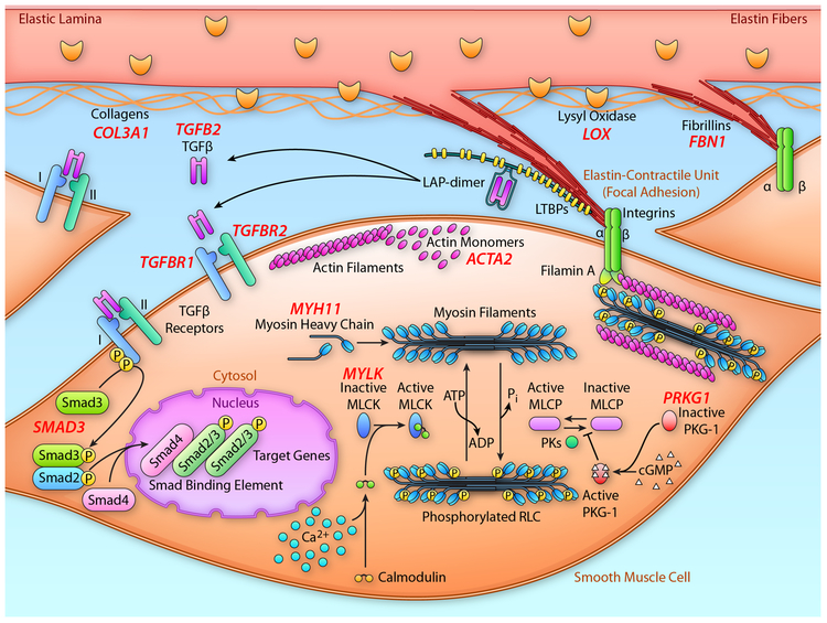
Figure 3:
Schematic representation elastin lamellae and SMCs highlighting the proteins that are disrupted by mutations in genes, leading to heritable thoracic aortic disease. Extensions from the elastin lamellae with fibrillin-1-containing microfibrils at the end link to integrin receptors on the cell surface of SMCs. The integrin receptors then link to the contractile filaments inside the cells, thus forming the elastin-contractile unit. Also illustrated is the proteins involved in canonical TGF-β signaling that are disrupted by mutations in the corresponding genes to also lead to heritable thoracic aortic disease. The validated genes predisposing to thoracic aortic disease are shown in red and are adjacent to their corresponding protein. MLCK; myosin light chain kinase; PKG-1, type I cGMP-dependent protein kinase. (Illustration Credit: Ben Smith).

