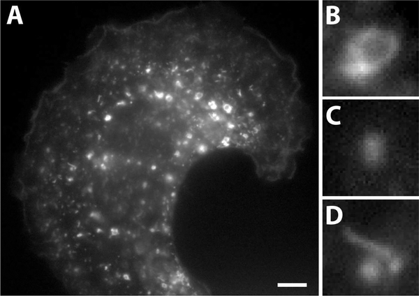
Fig. 1.
A microscopy image of endosomes in a cell and magnified images of representative structures. (A) A grayscale fluorescence microscopy image of a human myeloid endothelial cell expressing the fluorescently tagged marker protein, neonatal Fc Receptor (FcRn). (B) Ring-like endosomes. (C) Diffraction limited spots. (D) Tubule-like structures. Scale bar = 5 μm.

