


Figure 3.
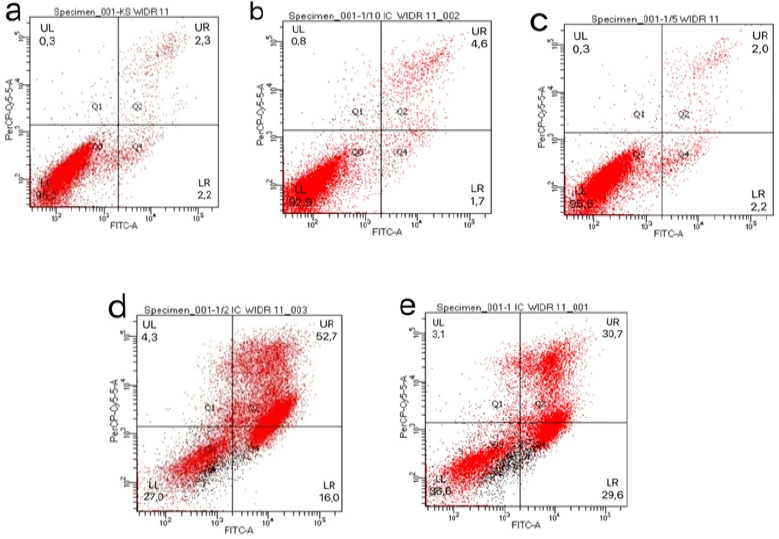
The Induction of Apoptosis in WiDr Cells by PNF. WiDr cells were treated with various concentrations of PNF for 24 h. The cells in the negative control group were untreated. The percentage of apoptotic cells was determined by annexin V/PI staining and flow cytometry. The cells generating an annexin V+/PI- (LR) signal were in the early apoptotic stage, while the cells generating an annexin V+/PI+(UR) signal were dead cells. Typical distribution of cell populations treated with 0/18/36/90/180 μg/mL PNF are shown in a-e, respectively.