


Figure 4.
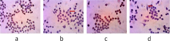
The Observation of Bcl-2 and Cyclin D1 Protein Expression in WiDr cells after Administration of PNF. The observations were made using an inverted microscope 400× magnification. (a), (c), The negative control, the group untreated with PNF; (b), cells incubated with PNF at a concentration of 90 µg/mL (1/2IC50) with antibodies against Bcl-2; (d), cells incubated with polyisoprenoids from N. fruticans at a concentration of 90 µg/mL (1/2 IC50) with antibodies against cyclin D1. The red arrow shows the negative result of Bcl-2 and cyclin D1 expression.