
Figure 3.
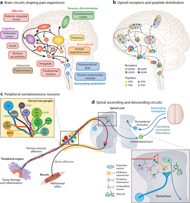
Neuroanatomical substrates of pain perception and remodeling by opioids. (a) A large interconnected neural network of supraspinal brain circuits transforms nociceptive information ascending from the spinal cord into an aversive, painful experience. (b) The opioid system is well positioned within this brain network to modify the perception of pain. The different opioid receptors and peptides are distinctively, though broadly, expressed in different sites, the function of which is under intense investigation. Relative opioid receptor (circles) and peptide (triangles) expression levels are denoted by the size of the shapes. (c,d) Opioid receptor types and peptides are also distributed in distinct subpopulations of (c) DRG neurons, identified with the indicated markers such as TRPV1, and (d) second-order spinal cord dorsal horn neurons. NF marks large-diameter DRG neurons with myelinated axons. Striped neurons coexpress different opioid receptor types. Abbreviations: CGRP, calcitonin gene-related peptide; DRG, dorsal root ganglion; DOPR, delta opioid receptor; DYN, dynorphin; END, p-endorphin; ENK, enkephalin; KOPR, kappa opioid receptor; MOPR, mu opioid receptor; MrgD, Mas-related G protein-coupled receptor member D; NF, neurofilament; NOC, nociceptin/orphanin FQ; NOPR, nociceptin opioid receptor; Ret, Ret proto-oncogene; TrkC, tropomyosin receptor kinase C; TRPV1, transient receptor potential cation channel subfamily V member 1.