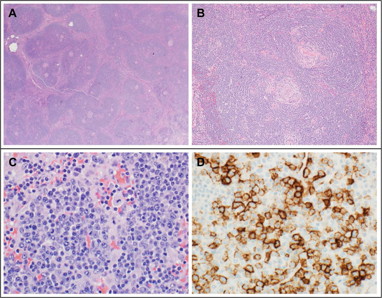
FIGURE 1. Representative histopathologic findings in Castleman disease.
(A) Hyaline vascular (HV) variant Castleman Disease (CD) in a 14 year old female with right neck mass: Low magnification reveals a broad mantle zone with several small and regressed germinal centers in lymphoid follicles. (B) HV disease (same patient): At a higher magnification, lymphoid follicles show a broad mantle zone with concentric rings (onion skin pattern) and small, regressed and hyalinized germinal centers radially penetrated by a blood vessel forming a lollipop pattern. (C) Plasma cell variant (PCV) CD in a 14 year old female with TAFRO (thrombocytopenia, anasarca, myelofibrosis, renal failure, and organomegaly) based on thrombocytopenia, anasarca, inflammation and lymphadenopathy: Lymph node biopsy reveals sheets of benign-appearing plasma cells. (D) PCV CD (same patient): Immunohistochemical stain CD138 highlights plasma cells.

