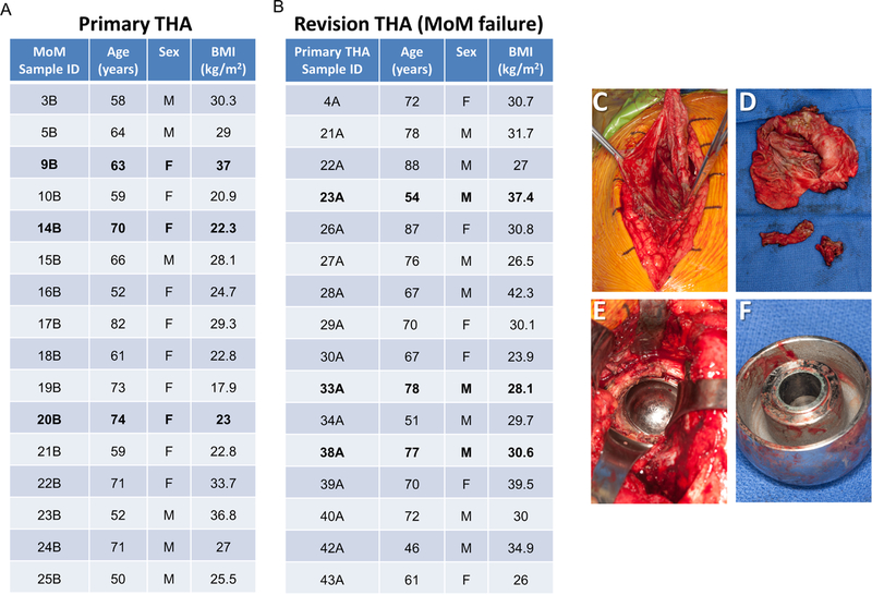
Figure 1.
Overview of the patient cohort and tissue biopsies used for RNA-seq (samples analyzed by RNA-seq are presented in bold font) (A-B). Surgical photograph of periacetabular tissue sampling in diseased patient (C) and surgical photograph of removed tissue sent for tissue processing and RNA-seq analysis (D). Intraoperative photograph of metal corrosion from metal-on-metal cobalt-chromium implant from acetabular component (E) and from explanted femoral head component (F) at the time of revision THA.

