FIG. 1.
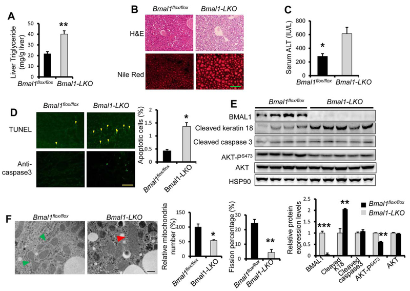
Hepatocyte-specific Bmal1 knockout mice are sensitive to ethanol feeding-induced liver steatosis and liver injury. Eight-week-old Bmal1Flox/Flox littermates and Bmal1Flox/Flox Alb-Cre(+) (Bmal1-LKO) mice were fed 5% ethanol diet for 10 days before binge feeding with ethanol (5 g/kg body weight) at Zeitgeber Time (ZT)3 and dissected 9 hours later (n 5 4 for Bmal1Flox/Flox and n = 5 for Bmal1-LKO, both male and females). (A) Hepatic triglyceride levels. (B) H&E staining and Nile red staining. (C) ALT assay to assess liver injury. (D) TUNEL staining and immune-fluorescence against cleaved caspase3 to assess apoptosis. (E) Western blotting analysis of hepatic apoptotic markers. (F) Transmission electron microscopy (magnification 40,000X). Mitochondria undergoing fission are indicated with green arrowheads and swollen mitochondrial with red arrowheads. *p < 0.05, **p < 0.01 by two-tailed Student’s t test. Scale bar = 100 μM for B and D; Scale bar = 400 nM for F.

