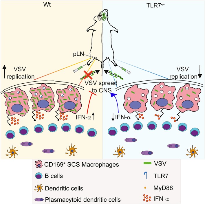
Figure 8.
Graphical abstract. (Left) VSV injected subcutaneously via foot of mice reaches to draining pLN where the viral particles are captured by CD169+ macrophages lining the subcapsular sinus (SCS) layer. Virus is recognized via TLR7 and promptly replicates in SCS macrophages due to their low type I IFN responsiveness and low levels of lysosomal enzymes. Replicating virus activates SCS macrophages which produce type-I IFN in situ and simultaneously trigger migration of pDC toward SCS layer where pDC encounter VSV and produce additional type-I IFN. High concentrations of type-I IFN prevent entry of virus in peripheral nerves. (Right) On the other hand, lymph-borne VSV captured by SCS macrophages inefficiently replicates in these cells lacking TLR7. Although SCS macrophages can secrete type-I IFN in a TLR7-independent manner, TLR7−/− pDC recruited to SCS layer cannot recognize VSV particles and so fail to produce type-I IFN. Impaired antiviral responses cause VSV dissemination into the central nervous system in the absence of TLR7 function.

