Figure 1.

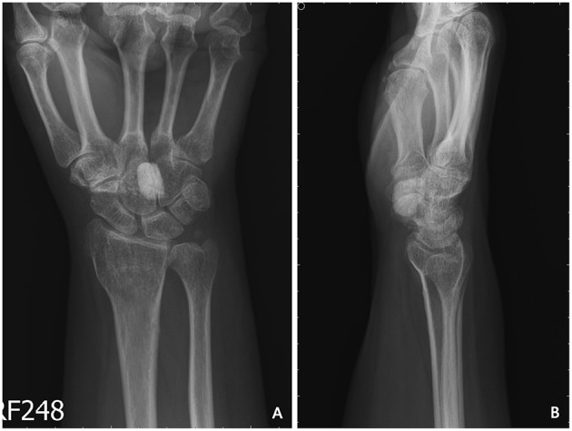
Preoperative X-ray of the first patient showing one calcified nodule sized 1.3 × 0.8 × 1.0 cm3 at the volar side of the capitate–hamate region. (A) Anteroposterior view. (B) Lateral view.

Preoperative X-ray of the first patient showing one calcified nodule sized 1.3 × 0.8 × 1.0 cm3 at the volar side of the capitate–hamate region. (A) Anteroposterior view. (B) Lateral view.