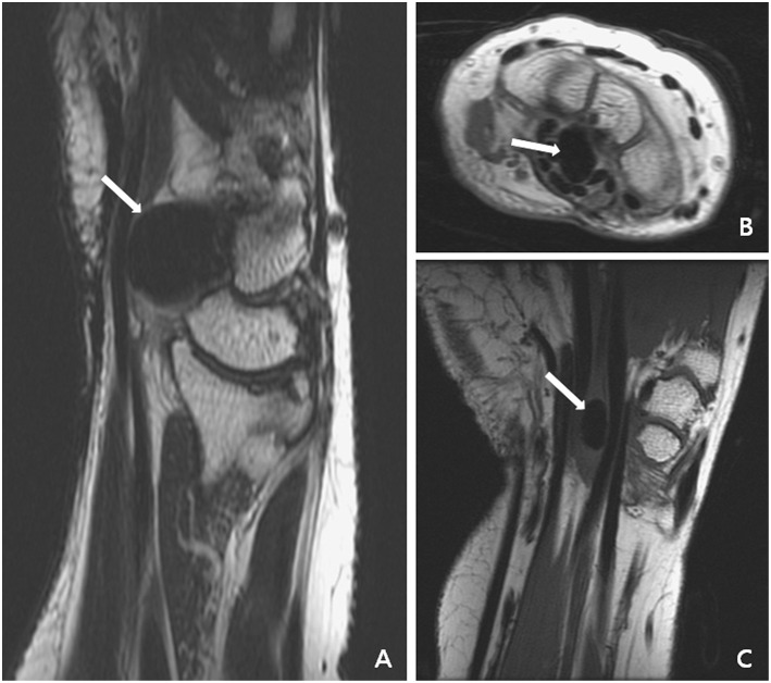
Figure 2.
Magnetic resonance imaging of the first patient disclosed focal lower intensity of the nodular lesion (white arrow) without obvious contrast enhancement. (A) Sagittal plane of T2- and T1-weighted images. (B) Axial plane of T1-weighted image with contrast. (C) Coronal plane of T1-weighted image.

