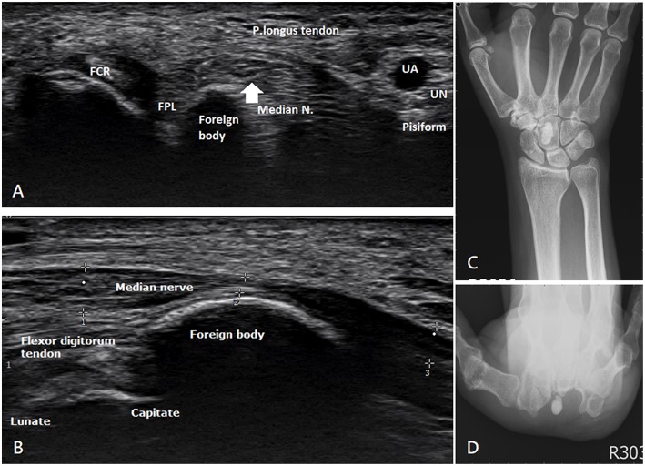
Figure 3.
Ultrasonography and preoperative X-ray of the second patient. (A) Transverse view and (B) longitudinal view present one hyperechoic ovoid lesion occupying the carpal tunnel with obvious median nerve compression. (C) Anteroposterior view and (D) carpal tunnel view show one radiopaque nodule sized 0.6 × 0.6 × 1.3 cm3 in front of the capitate.

