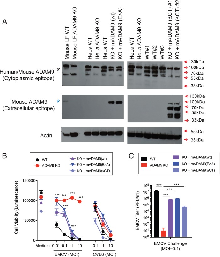
FIG 5.
Rescue of ADAM9 expression restores EMCV replication in ADAM9 KO cells. WT and ADAM9 KO HeLa cells were transduced with retroviral vectors with wild-type (WT) murine ADAM9 (mADAM9), catalytically inactive mutant ADAM9 (E>A), cytoplasmic-tail-deleted (ΔCT) ADAM9 constructs, or GFP control vectors. (A) WT, KO, and rescue cell lysates were analyzed by Western blot using two different ADAM9 antibodies. Top panel, rabbit anti-human ADAM9 that detects an epitope in the intracellular domain of human ADAM9 and cross-reacts with mouse ADAM9 (black asterisk). Middle panel, goat anti-mouse ADAM9 that detects the extracellular domain of murine ADAM9 but not human ADAM9 (blue asterisk). Bottom panel, anti-actin which detects both human and murine β-actin. Top panel, WT but not KO cells expressed human ADAM9. Middle panel, rescue but not KO cells expressed murine ADAM9. Bottom panel, actin loading control. (B) WT clones, ADAM9 KO clones, and rescued ADAM9-expressing clones were infected with EMCV or CVB3 at various MOIs and incubated at 37°C for 24 h. Viability of EMCV-infected and CVB3-infected clones was measured by CellGlo ATP luminescence. (C) EMCV replication was quantified in infected culture supernatants by plaque assay. Neither the functional sequence of the ADAM9 metalloproteinase domain nor the cytoplasmic tail is required for EMCV infection. ***, P < 0.0001, KO versus WT and KO versus rescue.

