FIG 7.
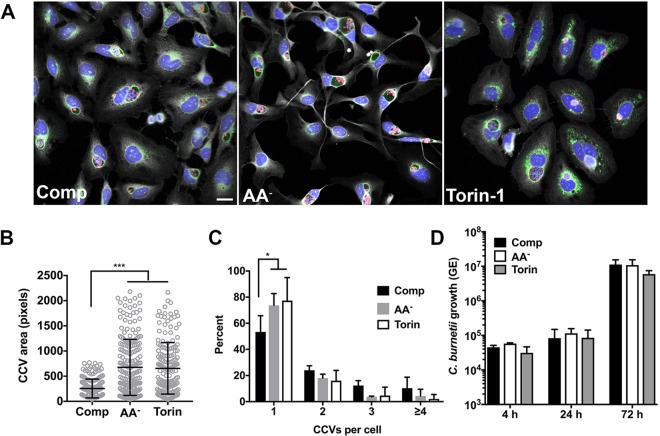
Autophagy promotes CCV expansion and fusogenicity without benefitting C. burnetii replication. (A) Representative fluorescence micrographs of infected HeLa cells incubated for 72 h in complete (Comp), AA−, or Torin-1 medium and then stained for DNA (blue), C. burnetii (red), LAMP1 (green), and cells (gray). (B) Quantitation of CCV size in infected HeLa cells incubated in Comp, AA−, or Torin-1 medium. The plot depicts means CCV size ± standard deviations (n = >100 cells) at 72 hpi. Data are representative of results from three independent experiments. (C) Quantitation of the number of CCVs per cell in cells incubated in Comp, AA−, or Torin-1 medium. The plot depicts mean percentages ± standard deviations from cells (n = >100) containing the indicated number of CCVs for three independent experiments. (D) Replication of C. burnetii in THP-1 macrophages incubated in complete, AA−, or Torin-1 medium. The plots depict means ± standard deviations of the fold change in bacterial genome equivalents (GE) relative to the mean at 4 hpi for three independent experiments. Asterisks indicate statistical significance (*, P < 0.05; ***, P < 0.001). Scale bar, 20 µm.

