Figure 2.

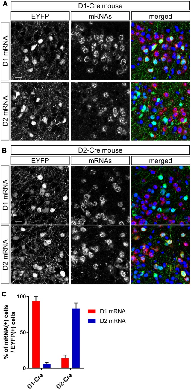
Cell type-specific gene expression in dopamine receptor D1- and D2-expressing neurons in the anteromedial OT of the D1-Cre and D2-Cre mice using AAV vectors. (A,B) Confocal images of AAV-derived EYFP-expressing cells (green) and D1 (upper panels) or D2 (lower panels) mRNAs (red) from a D1-Cre (A) or D2-Cre (B) mouse. Color merged panel contains DAPI staining (blue). Scale bars: 20 μm. (C) Percentage of D1 or D2 mRNA-expressing cells among EYFP-expressing cells in D1-Cre and D2-Cre mice. Data show mean with SD. OT, olfactory tubercle; AAV, adeno-associated virus; EYFP, enhanced yellow fluorescent protein; D1, dopamine receptor D1; D2, dopamine receptor D2; DAPI, 4′,6-diamidino-2-phenylindole; SD, standard deviation.
