Figure 4.

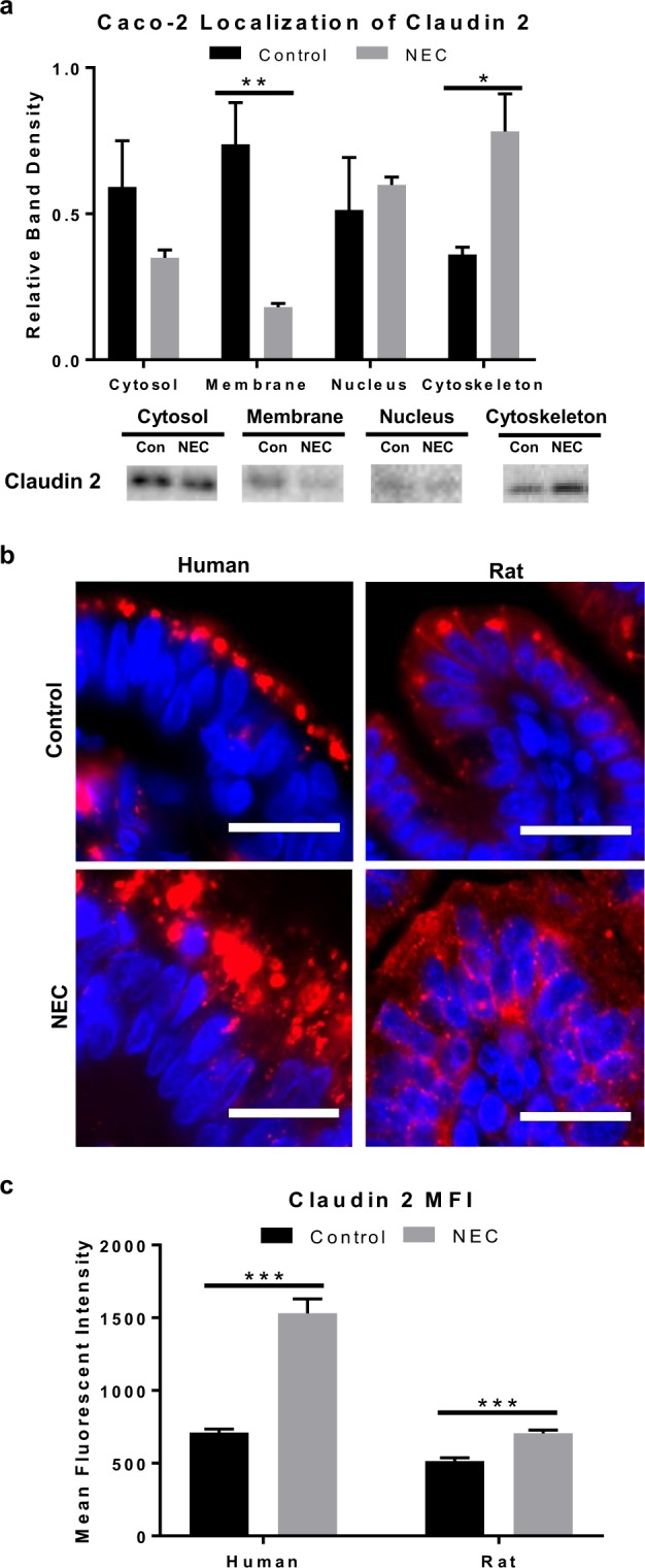
Human and experimental NEC is associated with changes in claudin 2 localization within the cell. (a) Caco-2 cells treated with LPS for 7 days were fractionated into subcellular compartments and analyzed with western blot showing a decrease in claudin 2 expression in the membrane of LPS-treated cells vs. controls (n = 5/4) and increased expression in the cytoskeleton vs. controls (n = 3/4); representative immunoblots for claudin 2 per compartment shown (full blots included in Supplementary Fig. S3). (b) Representative immunofluorescence micrographs of human and rat intestinal villi stained for claudin 2 (red) show focal expression at the cell membrane in control groups vs. internalized pattern of expression in NEC groups; nuclei stained with DAPI (blue), scale bar = 20 uM. (c) Mean fluorescent intensity measured in 15 × 30 um size boxes (2-cells wide) showed increased claudin 2 fluorescence in humans and rats NEC compared to controls (n = 3/3). All values are mean ± SEM (*p < 0.05, **p < 0.01, ***p < 0.001).
