Figure 5.
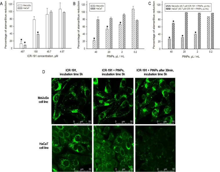
Influence of the platinum nanoparticles (PtNPs) on ICR-191 mutagenic activity in tested eukaryotic cell lines: (A) comparison of the cell viability of HaCaT and MelJuSo cell lines incubated for 72 h in a presence of ICR-191 (4.57–450 µM), based on alamarBlue assay. Data are expressed as the mean ± standard deviation and (B) comparison of the cell viability of HaCaT and MelJuSo cell lines incubated for 72 h in a presence of PtNPs (0.2–40 µL/mL), based on alamarBlue assay. Data are expressed as the mean ± standard deviation and (C) comparison of the cell viability of HaCaT and MelJuSo cell lines incubated for 72 h in a mixture of ICR-191 (45.7 µM) and PtNPs (0.2–40 µL/mL), based on alamarBlue assay. Data are expressed as the mean ± standard deviation, and (D) confocal microscopy live analysis of the impact of platinum nanoparticles (PtNPs) on ICR-191 fluorescence in the HaCaT and MelJuSo cell lines. Cells were treated with 45.7 µM ICR-191; treated with 45.7 µM ICR-191 and 3 ng/mL PtNPs mixture, or preincubated with 3 ng/mL PtNPs and subsequently treated with 45.7 µM ICR-191 (PtNPs preincubation). Time of incubation in the presence of ICR-191 indicated above particular panels. *Values significantly different from the amount of alamarBlue reduced by untreated control cells (p < α, α = 0.05).

