Figure 2.
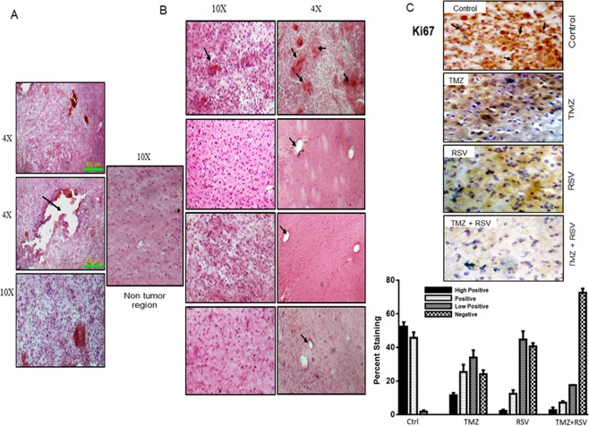
Roscovitine (RSV) alone or in combination with Temozolomide (TMZ) restricts glioma progression in vivo. (A) H and E staining of tumor sections on 7th day after tumor implantation was done in 2 randomly selected rats to confirm the development of tumor. Sections are shown at different magnification to show the spread of tumor from the site of injection (black arrow). (B) H and E staining of tumor sections (5–10 µ) from vehicle, TMZ, RSV and TMZ + RSV (from top to bottom) treated rats. After completion of the respective treatment cycles, rats were sacrificed. Brains were harvested and fixed in 4% PFA followed by sucrose gradient dehydration and cryosectioning. The sections thus obtained were used for H and E staining. Images were captured at 10 × and 4 × . Black arrows show the extensive presence of hemorrhages in vehicle treated tumors whereas hemorrhages were totally absent in all the treated tumors. (C) 25–30 µ sections were used for Immunohistochemical staining for Ki67 in tumor sections from vehicle, TMZ, RSV and TMZ + RSV treated rats. Black arrow shows clear nuclear staining for Ki67 in tumors from vehicle treated rats. Representative images of tumor sections are shown for each group.

