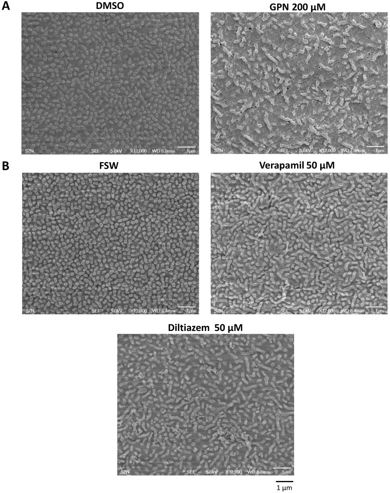
Figure 4.
Ultrastructural changes of the egg surface after the treatment with GPN, diltiazem and verapamil. (A) SEM images showing the microvilli of the unfertilized eggs treated with GPN (200 µM, 40 min) and DMSO. Note the reduced density of microvilli and their elongated shape in GPN-treated eggs in comparison with the control egg exhibiting regularly dispersed and shorter microvilli. (B) Effects of verapamil (50 µM, 40 min) and diltiazem (50 µM, 40 min) on the microvilli structure and quantity. Whereas SEM images of the control egg (FSW) display microvilli of regular distribution and length, the microvilli of the eggs treated with the two L-type Ca2+ channel inhibitors show irregular shapes and reduced quantity (also see Table 1). Scale bar 1 µm.

