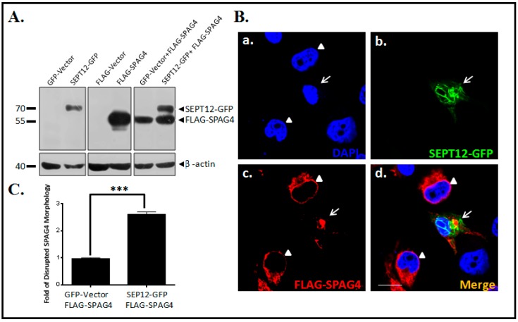
Figure 2.
SEPT12 overexpression disturbs SPAG4 localization around the NE in the NT2/D1 male germ cell line. (A) Western blot analysis of NT2D1 cells transfected with the pEGFP-empty vector (Lane1; GFP-Vector), pEGFP-SEPT12 (Lane2; SEPT12-GFP), pFLAGP-empty vector (Lane3; FLAG-Vector), pFLAG-SPAG4 (Lane4; FLAG-SPAG4), a mixture of the pEGFP-empty vector and pFLAG-SPAG4 vector (Lane5; GFP-Vector + FLAG-SPAG4), and a mixture of the pEGFP-SEPTIN12 vector and pFLAG-SPAG4 vector (Lane6; SEPT12-GFP + FLAG-SPAG4) using the anti-EGFP and anti-FLAG antibodies. (B) Immunofluorescence staining of NT2/D1 cells cotransfected with the pEGFP-SEPTIN12 vector and pFLAG-SPAG4 vector using DAPI (blue) (a), anti-EGFP antibody (green) (b) and anti-FLAG antibody (red) (c); (d) image obtained after merging the images in (a), (b), and (c). Magnification ×400 in (a–d). The arrow indicates the cells transfected with the pEGFP-SEPTIN12 vector (green) and pFLAG-SPAG4 vector (red). The arrowhead indicates the cells that were transfected only with the pFLAG-SPAG4 vector (red). Scale bar: 10 µm. (C) Quantification of the disorganization of SPAG4 in the NT2D1 cells transfected with a mixture of the pEGFP-empty vector and pFLAG-SPAG4 vector (Bar 1; GFP-Vector + FLAG-SPAG4) and a mixture of the pEGFP-SEPTIN12 vector and pFLAG-SPAG4 vector (Bar 2; SEPT12-GFP + FLAG-SPAG4). At least 100 transfected cells were counted in each experiment. Two-tailed Student t test; error bars indicate ± standard error of mean (*** p < 0.0001).

