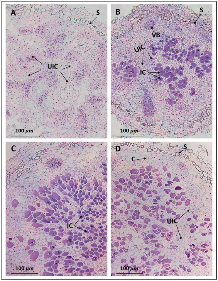
Figure 4.
Light micrographs of nodules. Pigeon pea nodules collected at 30 days after inoculation (DAI) were embedded in paraffin. Nodule sections were stained with hematoxylin and eosin. Note the nodules initiated by B. diazoefficiens USDA110 (A) contain only uninfected cells, while those inoculated with B. diazoefficiens Δ136 (B), S. fredii USDA191 (C), and S. fredii USDA RCB26 (D) revealed a distinct central infection zone. VB, vascular bundle; S, scleroid layer; UIC, uninfected cell; C, cortex.

