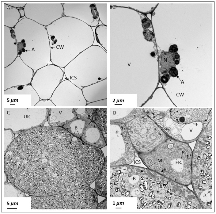
Figure 5.
Transmission electron micrographs of pigeon pea nodules. Thin sections of 30 DAI pigeon pea nodules were examined by transmission electron microscopy. Nodules elicited by B. diazoefficiens USDA110 contain no rhizobia inside the cells (A,B). These cells contain prominent vacuoles (V) that take up most of the cellular space (A,B). B. diazoefficiens Δ136-infected cells contain large number of bacteroids (C,D). CW, cell wall; ICS, intercellular space; M, mitochondria; ER, endoplasmic reticulum; N, nucleus; PBM, peribacteroid membrane; B, bacteroid; A, amyloplast; V, vacuole; UIC, uninfected cell.

