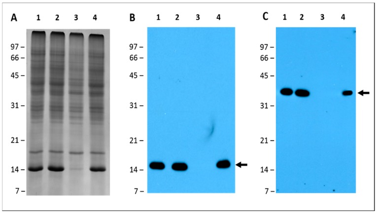
Figure 9.
Leghemoglobin and nitrogenase detection by immunoblot analysis. Three identical 15% SDS-PAGE gels were used to resolve pigeon pea total nodule proteins. One gel was visualized by staining with Coomassie Brilliant Blue (A) and the other two gels were electrophoretically transferred to a nitrocellulose membrane and probed with either the leghemoglobin antibody (B) or nitrogenase antibody (C). Immunoreactive proteins were detected using anti-rabbit IgG−horseradish peroxidase conjugate followed by chemiluminescent detection. The arrows point to the proteins reacting to the leghemoglobin and nitrogenase antibody, respectively. Lanes: 1, S. fredii USDA191 nodules; 2, S. fredii USDA RCB26 nodules; 3, B. diazoefficiens USDA110 nodules; 4, B. diazoefficiens Δ136 nodules. Molecular weight markers are shown on the left and designated in kDa.

