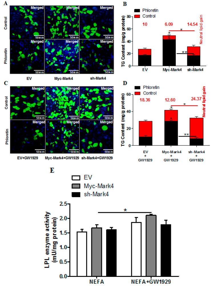
Figure 2.
MARK4 promotes lipid accumulation in pig primary trophoblast cells challenged with 400 μM NEFA. (A and C) Representative images (100×) of Bodipy staining after transfection with Myc-MARK4, sh-MARK4 for 48 h in primary (trophoblast cells) isolated from pig placentas. Primary trophoblasts were then incubated with 400 μM NEFA, 2 μM GW1929 or 500 μM phloretin for 24 h (n = 3). (B and D) Quantification of corresponding triglyceride (TG) in (A) and (C) by ELISA analysis (n = 3). The values in red indicate receptor (transport proteins)-mediated fatty acid accumulation by subtracting the values in the presence of phloretin from those in the absence of phloretin. (E) LPL activity (mU/mg protein) after transfection with Myc-MARK4, sh-MARK4 for 48 h in pig primary trophoblasts. Cells were then treated with 400 μM NEFA or 2 μM GW1929 for 24 h (n = 3). Values are expressed as mean ± SEM. ** p < 0.01; * p < 0.05 compared with the control group. Myc-MARK4 group: overexpression of MARK4 group, sh-MARK4 group: knock down of MARK4 group, Control: empty vector (EV) group.

