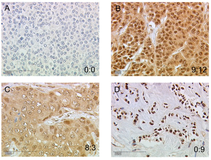
Figure 2.
Immunohistological staining of AhR in primary BC samples. AhR expression is illustrated in four tumors (A–D) with null or low nuclear expression (A,C) and high nuclear expression (B,D). The cytoplasmic:nuclear immunoreactive score (IRS) ratios are indicated for each sample. Scale bars: 100 μm.

