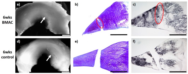
Figure 2.
(a) + (d): Macroscopic view of a lateral meniscus of New Zealand White rabbits. The created meniscus tears in the avascular parts of the pars intermedia are still visible in the control marked by the white arrow (d). In the bone marrow aspirate concentrate (BMAC)–meniscus suture group, macroscopically visible meniscus healing was found after 6 weeks, marked by the white arrow. (a). (b) + (e): Microscopic view (4× enlargement), DMMB staining of the lateral meniscus 6 weeks after surgery. No healing was seen in the control (e). Partial defect filling was observed 6 weeks after BMAC–meniscus suture treatment, marked with a red circle (b). (c) + (f): Microscopic view (4× enlargement). Collagen type II staining of the lateral meniscus 6 weeks after surgery. No healing was seen in the control (f). Partial defect filling was observed 6 weeks after BMAC–meniscus suture treatment as marked with a red circle (c). Benchmark: bar (white) = 2 mm, bar (black) = 1 mm.

