Figure 3.
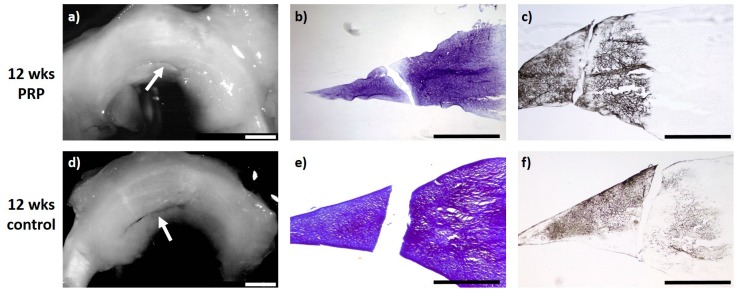
(a) + (d): Macroscopic view of a lateral meniscus of New Zealand White rabbits. The created meniscus tears in the avascular parts of the pars intermedia are still visible in the control (d) as well as the PRP–meniscus suture group (a) after a study period of 12 weeks, each marked by the white arrow. (b) + (e): Microscopic view (4× enlargement), DMMB staining of the lateral meniscus 12 weeks after surgery. No healing was seen in the control (e) or in the PRP–meniscus suture group (b). (c) + (f): Microscopic view (4× enlargement). Collagen type II staining of the lateral meniscus 12 weeks after surgery. No healing was seen in the control (f) or in the PRP–meniscus suture group (c). Benchmark: bar (white) = 2 mm, bar (black) = 1 mm.

